Rev Bras Fisiol Exerc 2022;21(2):125-34
doi: 10.33233/rbfex.v21i2.5068ORIGINAL ARTICLE
Cardiorespiratory Optimal Point application in
cardiopulmonary assessment in effort of individuals with spinal cord injury
Aplicação
do Ponto Ótimo Cardiorrespiratório na avaliação cardiorrespiratória em esforço
de indivíduos com lesão medular
Jeter Pereira de Freitas1, Míriam Raquel Meira Mainenti2, Camila Brasil e
Silva3, Patrícia dos Santos Vigário1
1Centro Universitário
Augusto Motta (UNISUAM), Rio de Janeiro, RJ, Brazil
2Escola
de Educação Física do Exército (EsEFEx), Rio de Janeiro,
RJ, Brazil
3Companhia de
Comando da 4a Brigada de Infantaria Leve de Montanha, Juiz de Fora, MG, Brazil
Recebido em: 27 de
janeiro de 2022; Aceito em: 23 de março de 2022.
Correspondência: Patrícia dos Santos Vigário, Programa de Pós-graduação
em Ciências da Reabilitação, Centro Universitário
Augusto Motta (PPGCR/ UNISUAM), Rua Dona Isabel, 94, Bonsucesso, 21041-020 Rio de Janeiro, RJ, Brazil
Jeter Pereira de Freitas: jeter.freitas@hotmail.com
Míriam Raquel Meira Mainenti:
miriam.mainenti@hotmail.com
Camila
Brasil e Silva: camilabrss@gmail.com
Patrícia
dos Santos Vigário: patriciavigario@yahoo.com.br
Abstract
Introduction: Spinal Cord
Injury (SCI) is related to low cardiorespiratory fitness and increased
cardiovascular morbidity and mortality. In individuals with SCI, the assessment
of cardiorespiratory capacity, whose best variable for analysis is maximal
oxygen consumption (VO2max), is commonly impaired due to early
interruption of effort. Consequently, measurements obtained at submaximal
intensities are necessary, such as the cardiorespiratory optimal point (POC). Objective:
To describe and compare the cardiorespiratory fitness in exertion of
individuals with high, low and no LM. Methods: Cross-sectional study in
participants with incomplete high LM, complete low LM and without LM, performed
with progressive tests on a cycle ergometer for upper limbs, considering peak
exercise, ventilatory threshold 1 (LV1) and POC. Results: Individuals
with SCI had lower exercise tolerance and lower peak VO2 compared to
individuals without SCI, despite the fact that all groups reached the end of
the exercise equally with a greater contribution of anaerobic metabolism in energy
production. As for the analysis of submaximal exertion intensities, individuals
with quadriplegia, among the three groups, reached maximum ventilatory
efficiency (POC) at higher percentages of peak VO2. Conclusion:
Individuals with SCI have lower cardiorespiratory fitness at peak and
submaximal exertion intensities when compared to individuals without SCI.
Particularly in relation to POC, the higher the level of LM, the greater the
ventilatory need to meet the metabolic demands of exercise.
Keywords: disabled persons; oxygen
consumption; exercise.
Resumo
Introdução: A Lesão Medular (LM) se relaciona à
baixa aptidão cardiorrespiratória e ao aumento da morbimortalidade
cardiovascular. Em indivíduos com LM, a avaliação da capacidade
cardiorrespiratória, cuja melhor variável para análise é o consumo máximo de
oxigênio (VO2máx), é comumente prejudicada devido à interrupção
precoce do esforço. Consequentemente, as mensurações obtidas em intensidades
submáximas se fazem necessárias, como o ponto ótimo cardiorrespiratório (POC). Objetivo:
Descrever e comparar a aptidão cardiorrespiratória em esforço de indivíduos com
LM alta, baixa e sem LM. Métodos: Estudo seccional em participantes com LM alta
incompleta, LM baixa completa e sem LM, realizado com testes progressivos em cicloergômetro para membros superiores, considerando pico
do exercício, limiar ventilatório 1 (LV1) e POC. Resultados: Os
indivíduos com LM apresentaram menor tolerância ao esforço e menor VO2
de pico em relação aos indivíduos sem LM, apesar de todos os grupos terem
chegado igualmente ao término do exercício com uma maior contribuição do
metabolismo anaeróbio na produção de energia. Quanto às análises em
intensidades submáximas de esforço, os indivíduos com tetraplegia, dentre os
três grupos, foram aqueles que alcançaram a máxima eficiência ventilatória
(POC) em percentuais mais altos do VO2 de pico. Conclusão:
Indivíduos com LM apresentam menor aptidão cardiorrespiratória no pico e em
intensidades submáximas de esforço quando comparados com indivíduos sem LM.
Particularmente em relação ao POC, quanto mais alto o nível da LM, maior a
necessidade ventilatória para o atendimento das demandas metabólicas do
exercício.
Palavras-chave: pessoas com deficiência; consumo de
oxigênio; exercício físico.
Introduction
Spinal cord injury (SCI) is
associated with changes in the functioning of several body systems, which may
vary according to the height of the injury [1]. In parallel, a higher
prevalence of sedentary behavior is described in individuals with SCI due to
environmental barriers - such as lack of accessibility - and psycho-emotional
barriers, such as demotivation and impaired self-esteem and self-image [2].
Together, these factors contribute to poor cardiorespiratory fitness and
increase the risk of health-related complications [3].
Maximum oxygen consumption (VO2max)
is the variable that best describes the cardiorespiratory fitness of
individuals [4], and higher values are related to a lower risk of
cardiovascular morbidity and mortality [5]. It is obtained through the
metabolic analysis of ventilatory gases during the performance of a maximum
progressive effort, commonly in cycle ergometers selected according to the
individual characteristics of the evaluated. In individuals with functional
limitations, as in SCI, however, it is common to interrupt the effort due to
peripheral factors such as low muscle capacity, limiting the performance of the
movement [6]. This makes it difficult to obtain the VO2max, limiting
the interpretation of the test results [7]. Faced with this difficulty, Ramos et
al. [8] proposed the analysis of the lowest value of the ventilatory oxygen
equivalent during exercise as an indicator that would represent the highest
ventilatory savings for the capture of oxygen and consequent supply to the
active musculature. This variable was named “Cardiorespiratory Optimal Point”
(COP), with the advantage of obtaining it at submaximal exertion intensity. It
is an alternative variable for the analysis of cardiorespiratory capacity,
especially in situations where maximum effort is not reached (as in different
functional limitations) or is not desirable (as in certain phases of sports
training) [9]. After consulting the Pubmed/Medline
and Scielo databases using the combination of the
“cardiorespiratory optimal point” and “spinal cord injury” terms, no studies
were found that had analyzed this variable in the population of individuals
with SCI.
The importance of a periodic
assessment of the cardiorespiratory capacity of individuals with SCI lies in
identifying and acting in different scenarios, including rehabilitation and
physical training prescription for sports purposes [10]. Therefore, it is
necessary that the measured variables effectively reproduce the reality and the
level of physical conditioning so that efficient interventions are selected and
the gains maximized. Thus, the objective of the present study is to test the
hypotheses: a) individuals with SCI have lower cardiorespiratory fitness than
individuals without SCI at different intensities of effort, and b) specifically
concerning COP, individuals with SCI have a greater ventilatory need during the
exercise.
Methods
Study and participants
A cross-sectional comparative
observational study was carried out with the participation of 27 men aged 18
years or over divided into three groups: incomplete high SCI (QUADRI group;
from the fourth to the seventh cervical vertebra; N = 09), complete low SCI
(PARA group; first thoracic vertebra to second lumbar vertebra; N = 08) and without
SCI (N = 10). All were physically active for at least six months: wheelchair
rugby practice in the QUADRI group, wheelchair basketball in the PARA group,
and aerobic and strength exercises in the group without SCI. Individuals
without SCI were classified as “active” or “very active” by completing the
short version of the International Physical Activity Questionnaire (IPAQ) [11].
The groups of participants with SCI were selected for convenience in two sports
associations for people with disabilities in Rio de Janeiro, Brazil. Smokers,
users of substances that interfere with the heart rate response (for example,
beta-blockers, sympathomimetics, and sympatholytics),
and those with pain or disabling musculoskeletal limitations for performing the
Cardiopulmonary Exercise Test (CPET) were excluded.
The study was approved by the
institutional Research Ethics Committee (CAAE: 37041520.4.0000.5235), and all
participants signed an informed consent form before participating in the study.
Cardiorespiratory fitness in exertion
To assess cardiorespiratory fitness
during exercise, a CPET of increasing intensity was performed on a cycle
ergometer for the upper limbs (TopExcite, TechnoGym; Italy). The tests were performed in the morning
in a laboratory with controlled temperature (≈ 22o C) and
humidity (≈ 60%) [12].
The initial load was 20w with
successive increments of 2w or 5w every minute – according to the functionality
of the participant's upper limbs – and cycling between 50-60 rpm [13].
Participants were verbally encouraged to exert maximum effort, which was
interrupted due to exhaustion or the appearance of one of the criteria defined
by the American College of Sports Medicine (2018) [4].
Throughout the test, the
participants remained connected to a metabolic analyzer of ventilatory gases
(VO2000, MedGraphics; Brazil) that allowed the
reading of pulmonary ventilation (VE, L/min) and expired oxygen fractions (FeO2,
%) and carbon dioxide (FeCO2, %). Information was recorded
breath-by-breath and plotted as a 30-second average. The following variables
were calculated: relative and absolute oxygen consumption (VO2,
mL.kg-1.min-1, and L/min, respectively) and ventilatory
equivalents of oxygen (VE/VO2) and carbon dioxide (VE/VCO2).
VO2 was considered maximum if: (I) presence of a plateau in the VO2
curve concomitant with the increase in effort intensity was observed; (II)
respiratory quotient (R) ≥ 1.1; and (III) existence of VT1 [12]. In the
absence of VO2max, the highest value presented in the last minute of
the test was considered as peak.
Ventilatory threshold 1
Ventilatory threshold 1 (VT1) was
identified according to the recommendations proposed by Gaskill et al.
[14], which include the combination of analysis of three methods: I) ventilatory
equivalents of oxygen and carbon dioxide; II) excess carbon dioxide; and III)
modified V-slope. Visual inspection to determine VT1 was performed
independently by two experienced evaluators. If the difference between the
evaluators concerning VO2 in VT1 was within 3%, the mean value was
adopted as the final result. If the difference exceeded 3%, a third evaluator
would be asked to determine VT1.
Cardiorespiratory Optimal Point
The Cardiorespiratory Optimal Point
(COP) was considered the lowest value of the ventilatory oxygen equivalent
(VE/VO2) during the effort [8]. Relative VO2 (mL.kg-1.min-1),
absolute VO2 (L/min), percentage of peak VO2, load (w),
and effort time (s) relative to COP were also analyzed.
Statistical procedures
Since the eligible sample consisted
of all wheelchair rugby athletes from a regional team, a post-hoc calculation
of the minimum effect size (f) to be detected was performed for comparison
between groups using the G*Power software. Considering a type I error equal to
5% and test power equal to 80% (type II error equal to 20%), a minimum effect
size (f) of 0.63 can be detected in the 3 groups comparison, with a total of 27
participants.
The exploratory data analysis was
presented by the median and the minimum and maximum values. The distribution of
variables was verified using the Shapiro-Wilk test, and after the analysis, it
was decided to adopt non-parametric procedures. Comparisons between the three
subgroups of the study were made using the Kruskal-Wallis test and the
differences identified by the Mann-Whitney test with Bonferroni correction for
the three possible combinations of pairs (p < 0.017). Comparisons between
the QUADRI and PARA groups were made using the Mann-Whitney test. The statistical
significance level adopted was 5%, and the analyzes were performed using SPSS
20.0 (Armonk, NY: International Business Machines Corporation).
Results
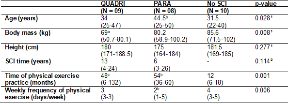
The general characteristics and the
practice of physical exercises of the study participants are described in Table
I. In comparison with the group without SCI, the QUADRI group had lower body
mass, and the PARA group was older. The time of physical exercise practice was
similar between the QUADRI and PARA groups and longer than that presented by the
group without SCI. There was also no difference between the time of SCI.
Table I - General characteristics of
the study participants, according to the analysis subgroup

QUADRI = High and incomplete spinal
cord injury; PARA = low spinal cord injury; SCI = spinal cord injury;
1Kruskal-Wallis test, statistical significance when p < 0.05; ²Mann-Whitney
test, statistical significance when p < 0.05; aMann-Whitney
test with Bonferroni correction, statistical significance when p < 0.017
(QUADRI ≠ PARA); bMann-Whitney test with
Bonferroni correction, statistical significance when p < 0.017 (PARA ≠
Without SCI); c Mann-Whitney test with Bonferroni correction, statistical
significance when p < 0.017 (QUADRI ≠ Without SCI)
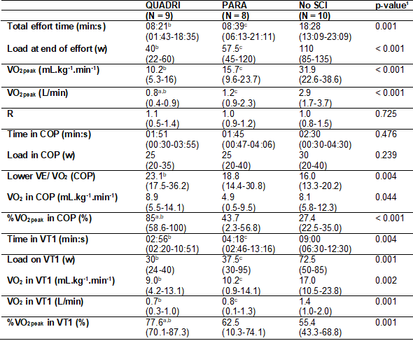
Table II shows the results related
to CPET. The QUADRI and PARA groups presented lower time and total effort load,
relative VO2peak, and absolute VO2peak, compared to the
group without SCI. The R at the end of the effort, however, did not differ
between the three groups. All participants reported interruption of effort due
to peripheral fatigue of the upper limbs involved in the movement.
Regarding the COP, the groups
showed similarity in terms of time at the moment of reaching (p = 0.476) and
load (p = 0.239). The COP value was lower in the QUADRI group compared to those
without SCI and the %VO2peak in the COP was higher in the QUADRI
both in relation to the PARA and in relation to those without SCI. One
participant in the QUADRI group presented COP after VT1 (COP time = 03 min:
52s; VT1 time = 02 min: 51s). All other participants presented COP before VT1.
Of all study participants, only two in the QUADRI group did not reach VT1. The
QUADRI and PARA groups reached this point earlier than those without SCI, as
well as with lower loads, VO2 (mL.kg-1.min-1)
and VO2 (L/min). The %VO2peak in VT1 was higher in the
participants of the QUADRI group in relation to the PARA and without SCI.
Table II – Variables related to the
cardiopulmonary exercise test of the study participants, according to the
analysis subgroup

QUADRI = High and incomplete spinal
cord injury; PARA = low spinal cord injury; SCI = spinal cord injury;
1Kruskal-Wallis test, statistical significance when p < 0.05; Mann-Whitney
test with Bonferroni correction, statistical significance when p < 0.017
(QUADRI ≠ PARA); bMann-Whitney test with
Bonferroni correction, statistical significance when p < 0.017 (QUADRI ≠
Without SCI); cMann-Whitney test with Bonferroni
correction, statistical significance when p < 0.017 (PARA ≠ Without SCI)
Discussion
The main findings of the present
study include lower exercise tolerance and lower peak VO2 in
participants with SCI compared to individuals without SCI, even though all
groups reached the end of the exercise equally with a higher contribution of
anaerobic metabolism in the process of energy production (mean R of the groups ≥
1; p = 0.725). About the analysis of submaximal exertion intensities,
individuals with quadriplegia reached maximum ventilatory efficiency, that is,
COP, at higher percentages of peak VO2, as well as VT1. This
difference was observed both in comparison to individuals with paraplegia and
those without SCI. To the authors' knowledge, this was the first approach to
the COP application in individuals with SCI.
The lower effort tolerance observed
in the SCI group may reflect the lower amount of muscle mass generally
presented in the upper limbs of these individuals, especially those with higher
injuries in which the loss is more pronounced. These structural losses impact
functionality and mobility by the total or partial interruption of sensory and
motor information below the lesion level [1]. However, it should be considered
that the exercise was performed with the upper limbs, which are highly
requested by individuals with SCI for daily commuting, but the movement
performed to propel the wheelchair differs from the gesture performed on the
cycle ergometer used. Therefore, it is also possible that this difference
influenced, in a certain way, the occurrence of early fatigue.
As for VO2 at the end of
the exercise, the differences observed between individuals with and without
SCI, in addition to being explained by the amount of muscle mass involved
during the test, may also be associated with the modulation of cardiac
inotropism by the autonomic nervous system [15]. Through the sympathetic and
parasympathetic branches, which, respectively, increase and decrease cardiac
function, changes in heart rate and contraction force are observed, for example
[16]. Since the sympathetic fibers that innervate the heart originate in the
spinal cord, between the first and fifth thoracic vertebrae, and the
parasympathetic fibers originate in the vagus nerve,
it is concluded that individuals with SCI at these levels have impairments in
sympathetic tone and, consequently, in the positive stimulus to cardiac work.
According to Draghici and Taylor [17], the damage to
the autonomic pathways in the face of an SCI may not be necessarily associated
with the level/height of the lesion, or even if it is complete or incomplete.
However, considering the origins of the sympathetic and parasympathetic fibers
that innervate the heart, the sympathetic control would be more impaired the
higher the level/height of the lesion, while the parasympathetic control would
remain unchanged.
Impairments in sympathetic control
include lower heart rate at submaximal and maximal exertion levels, lower blood
pressure, and lower cardiac output. These changes during physical exercise
translate into a lower blood supply to the active muscles and, consequently,
lower VO2 [18]. Nightingale et al. [19] investigated the
cardiorespiratory capacity in effort on a cycle ergometer for upper limbs of
patients with cervical SCI, high thoracic SCI, and thoracolumbar SCI, noting
differences between the groups both with peak VO2 and maximal power
- that is, how much the higher the lesion height/level, the lower the VO2
and power. In a study comparing cardiorespiratory capacity under two conditions
a) using only the cycle ergometer for upper limbs and b) the cycle ergometer
associated with electrical stimulation, an inverse relationship was also
observed between the height/level of the lesion and, VO2peak
regardless of the condition tested [20]. It is noteworthy that, in the presence
of electrical stimulation, the VO2 values were higher. This same
relationship between SCI level/height was observed in the present study,
although without statistical significance in some comparisons.
In the measurements obtained, the
COP was one of the moments considered for the cardiorespiratory capacity
characterization in submaximal effort intensity. In the study by Ramos and
Araújo [21], COP values lower than 22 were shown to be associated with a lower
risk of mortality both in healthy individuals and in individuals with chronic
diseases. However, it should be noted that the assessment was performed on a
cycle ergometer for lower limbs, and this value will not necessarily be the
same for exercises performed on other ergometers, such as the treadmill and
cycle ergometer for upper limbs. To date, the authors are not aware of a
reference point delimitation for the mortality risk classification by analyzing
the COP obtained using a cycle ergometer for upper limbs. However, if lower COP
values are associated with a lower risk, it can be assumed, in the present
study, that the QUADRI group may be at greater risk or, more appropriately,
present a worse ventilatory economy since it presented the highest Median COP
among the three subgroups evaluated in the study (median value of the QUADRI
group = 23.1). Therefore, for individuals with higher SCI, a greater volume of
air would be necessary to be ventilated for the consumption of 100 mL of O2
for individuals with low SCI and individuals without SCI. Concerning
individuals with low SCI, this same analysis is reproduced about individuals
without SCI.
Another observation that
corroborates the lower cardiorespiratory capacity of the QUADRI group
concerning the other groups is the percentage of VO2peak at which
the COP was reached. Even with reference values for cycle ergometers other than
the one for upper limbs, the literature indicates that the COP is reached, on
average, between 30 and 50% of VO2max/peak [8]. The referred group
reached the COP with about 85% of the peak VO2 (median value), that
is, closer to the highest value of O2 consumed during the test. This may
suggest that after reaching the greatest ventilatory economy, there was not a
very expressive increase in VO2 until the end of the test.
VT1, which represents the beginning
of the transition from the predominance of aerobic to anaerobic metabolism, was
another submaximal measure in which individuals with SCI presented lower
performance than those without SCI. The earlier reaching of this point may be
associated with the same factors discussed above; lower muscle mass in the
upper limbs, a motor gesture performed in the test different from the one
usually used in day-to-day wheelchair propulsion, and changes in cardiac
autonomic control. It is noteworthy that, even if individuals with SCI have
reached VT1 in higher percentages of VO2peak compared to individuals
without SCI, this does not reflect better cardiorespiratory fitness since the
VO2peak of these individuals was, in general, low.
A proposal to analyze the
aforementioned variables was recently carried out by Costa et al. [22]
in patients with functional limitation due to unilateral lower limb amputation,
a condition that causes mobility impairment and less tendency to exercise, as
seen in SCI. In the individuals tested when compared with individuals without
amputation, it was also verified lower values of VO2peak for the
same intensity of effort; and the COP reached higher percentages of VO2peak.
The lower cardiovascular performance of individuals with amputations, similarly
to the results of the present study, suggests that mobility limitations,
associated with the daily difficulties they impose, may constitute a risk
factor for lower cardiovascular capacity.
The present study has as a
limitation the performance of CPET on a cycle ergometer for upper limbs and not
on a treadmill adapted for a wheelchair in the case of individuals with SCI.
Thus, the daily motor gesture could be reproduced, reflecting more accurately
the cardiorespiratory capacity of these subjects. However, this is still an
unprecedented investigation in this population, which may benefit from the
prescription of physical exercises and rehabilitation more specifically
indicated for targeted functional gains. There is also the understanding that
the analyzed sample belongs to a minority of patients with SCI, considering
that a large part of these individuals does not have access to the regular
practice of adapted exercises. In future studies, it is suggested to approach
sedentary participants, especially considering the significant prevalence of
individuals with SCI and the increased cardiovascular risk in this population,
and thus contribute to more concretely establishing this risk and individualizing
approaches to minimize it.
Conclusion
Individuals with SCI have lower
cardiorespiratory fitness at peak and submaximal exertion intensities when
compared to individuals without SCI. Concerning COP, the higher the level of
SCI, the greater the ventilatory need to meet the metabolic demands of
exercise.
Conflict of interest
No potential conflicts of interest
relevant to this article have been reported.
Funding source
This study was partially funded by
the Coordination for the Improvement of Higher Education Personnel – Brazil
(CAPES) – Funding code 001, by the Fundação de Amparo
à Pesquisa do Estado do Rio de Janeiro (FAPERJ)
(public notice E-26/203.256/2017) and by the National Council for Scientific
and Technological Development (CNPq). The authors
also thank the Brazilian Paralympic Academy, of the Brazilian Paralympic
Committee (APB/CPB), for their scientific support.
Authors' contributions
Research conception and
design: Freitas JP, Vigário PS, Mainenti MRM; Data collection: Freitas JP; Data analysis
and interpretation: Freitas JP, Vigário PS, Mainenti MRM; Statistical analysis: Vigário PS, Mainenti MRM; Obtaining
financing: Vigário PS; Writing of the manuscript:
Freitas JP, Vigário PS, Mainenti
MRM; Critical review of the manuscript for important intellectual content:
Silva CB
References
- Raguindin PF, Bertolo
A, Zeh RM, Fränkl G, Itodo OA, Capossela S, et al. Body
composition according to spinal cord injury level: a systematic review and
meta-analysis. J Clin Med 2021;10(17):3911. doi: 10.3390/jcm10173911 [Crossref]
- Verschuren O, Dekker B, van Koppenhagen C, Post M. Sedentary behavior in people with spinal cord injury. Arch Phys Med Rehabil 2016;97(1):173. doi: 10.1016/j.apmr.2015.10.090 [Crossref]
- Maher JL, McMillan DW, Nash MS. Exercise and health-related risks of physical deconditioning after spinal cord injury. Top Spinal Cord Inj Rehabil 2017;23(3):175-187. doi: 10.1310/sci2303-175 [Crossref]
- ACSM.
Diretrizes do ACSM para os testes de esforço e sua prescrição. 10 ed. Rio de
Janeiro: Guanabara Koogan; 2018.
- Khan H, Jaffar N, Rauramaa R, Kurl S, Savonen K, Laukkanen JA. Cardiorespiratory fitness and non fatal cardiovascular events: A population-based follow-up study. Am Heart J 2017;184:55-61. doi: 10.1016/j.ahj.2016.10.019 [Crossref]
- Bento S,
Carvalho MP, Faria F. Recondicionamento ao esforço na lesão medular. Revista da SPMFR 2016; 28(1):22-28.
- American Thoracic Society; American College of Chest
Physicians. ATS/ACCP Statement on cardiopulmonary exercise testing. Am J Respir
Crit Care Med 2003;167(2):211-77. doi: 10.1164/rccm.167.2.211 [Crossref]
- Ramos PS, Ricardo DR, Araújo CGS. Cardiorespiratory optimal point: A submaximal variable of the Cardiopulmonary Exercise Testing. Arq Bras Cardiol 2012;99(5):988-96. doi: 10.1371/journal.pone.0104932 [Crossref]
- Silva CGS, Castro CLB, Franca JF, Bottino A, Myers J, Araújo CGS. Ponto ótimo cardiorrespiratório em futebolistas profissionais: Uma nova variável submáxima do exercício. Int J Cardiovasc Sci 2018;31(4):323-32. doi: 10.5935/2359-4802.20180030 [Crossref]
- Mercier H, Taylor JA. The physiology of exercise in
spinal cord injury (sci): an overview of the limitations and adaptations. The
Physiology of Exercise in Spinal Cord Injury 2016;1-11 10. doi: 10.1007/978-1-4939-6664-6_1 [Crossref]
- Matsudo S, Araújo T, Matsudo V, Andrade D, Andrade E, Oliveira LC, et al. Questionário Internacional de Atividade Física (IPAQ): estudo de validade e reprodutibilidade no Brasil. Rev Bras Ativ Fís Saúde 2012;6(2):5-18. doi: 10.12820/rbafs.v.6n2p5-18 [Crossref]
- Yazbek PJ, Carvalho RT, Sabbag LMS, Battistella LR. Ergoespirometria. Teste de esforço cardiopulmonar, metodologia e interpretação. Arq Bras Cardiol 1998;71(5):719-24. doi: 10.1590/s0066-782x1998001100014 [Crossref]
- Campos LFCC. Comparação entre métodos para mensuração da potência aeróbia em atletas tetraplégicos [Dissertação]. São Paulo: Faculdade de Educação Física da UNICAMP; 2013. doi: 10.47749/T/UNICAMP.2013.902347 [Crossref]
- Gaskill SE, Ruby BC, Walker AJ, Sanchez OA, Serfass RC, Leon AS. Validity and reliability of combining
three methods to determine ventilatory threshold. Med Sci Sports Exerc 2001;33(11):1841-8. doi: 10.1097/00005768-200111000-00007 [Crossref]
- Biering-Sørensen F, Biering-Sørensen T, Liu N, Malmqvist L, Wecht JM, Krassioukov A. Alterations in cardiac autonomic control in spinal cord injury. Auton Neurosci 2018;209:4-18. doi: 10.1016/j.autneu.2017.02.004 [Crossref]
- Herring N, Kalla M, Paterson
DJ. The autonomic nervous system and cardiac arrhythmias: current concepts and
emerging therapies. Nat Rev Cardiol 2019;16(12):707-26. doi: 10.1038/s41569-019-0221-2 [Crossref]
- Draghici AE,
Taylor JA. Baroreflex autonomic control in human spinal cord injury:
physiology, measurement, and potential alterations. Auton
Neurosci 2018;209:37-42. doi: 10.1016/j.autneu.2017.08.007 [Crossref]
- Gee CM, West CR, Krassioukov
AV. Boosting in elite athletes with spinal cord injury: A critical review of
physiology and testing procedures. Sports Med 2015; 45(8):1133-42. doi: 10.1007/s40279-015-0340-9 [Crossref]
- Nightingale TE, Bhangu GS, Bilzon JLJ, Krassioukov AV. A cross-sectional comparison between cardiorespiratory fitness, level of lesion and red blood cell distribution width in adults with chronic spinal cord injury. J Sci Med Sport 2020; 23(2):106-11. doi: 10.1016/j.jsams.2019.08.015 [Crossref]
- Shaffer RF, Picard G, Taylor JA. Relationship of
spinal cord injury level and duration to peak aerobic capacity with arms-only
and hybrid functional electrical stimulation rowing. Am J Phys Med Rehabil 2018;97(7):488-91. doi: 10.1097/PHM.0000000000000903 [Crossref]
- Ramos PS, Araújo CGS. Cardiorespiratory optimal point
during exercise testing as a predictor of all-cause mortality. Rev Port Cardiol 2017;36(4):261-9. doi: 10.1016/j.repce.2016.09.011 [Crossref]
- Costa RMR, Lisboa PMA, Santos MA, Mainenti MRM, Lopes AJ, Vigário PS. Aptidão cardiorrespiratória durante o teste cardiopulmonar de esforço de indivíduos com amputação unilateral de membro inferior. Rev Bras Fisiol Exerc 2021;20(5):542-551. doi: 10.33233/rbfex.v20i5.4824 [Crossref]
