Rev
Bras Fisiol Exerc. 2025;24:e245633
REVIEW
Exercise-induced myokines and their role in central
nervous system function: a scoping review
Miocinas induzidas pelo
exercício físico e suas funções no sistema nervoso central: uma revisão de
escopo
Pedro Henrique Sobral Neves¹, Paula Araujo2
1.
Escola Bahiana de Medicina e Saúde Pública, Salvador, Bahia, Brazil
2.
Universidade
Federal da Bahia, Bahia, Brazil
Received:
May 13, 2025; Accepted: September 9, 2025.
Correspondence: Paula Araujo,
E-mail: araujo.pca@gmail.com
How to
cite
Neves PHS, Paula Araujo P. Exercise-induced myokines and their role in central nervous system function:
a scoping review. Rev Bras Fisiol
Exerc 2025;24(1):e245633. doi: 10.33233/rbfex.v24i1.5633
Abstract
Introduction: The increasing prevalence of neurological diseases is
a major global concern. Recently, myokines, molecules secreted by active
skeletal muscle, have emerged as potential mediators of the beneficial effects
of physical exercise on the central nervous system (CNS), playing a key role in
muscle-brain communication. Objective:
To
review the scientific evidence on the influence of
exercise-induced myokines on central nervous system function. Methods: This scoping review
included clinical trials evaluating the secretion of exercise-induced myokines
and their impact on CNS function in healthy individuals and patients with
neurological disorders. Literature searches were conducted in PubMed/Medline, SciELO, and Embase, without time restrictions. Results: Eleven studies met the
inclusion criteria. Exercise protocols included aerobic training (seven
studies) and strength training (four studies), involving participants aged
23–69 years with or without CNS dysfunctions such as amnesia, Alzheimer’s
disease, Parkinson’s disease, and Prader-Willi syndrome. The most investigated
myokines were irisin and brain-derived neurotrophic factor (BDNF), although
findings were heterogeneous. Some studies demonstrated increased levels of
these myokines associated with improvements in cognitive function and balance,
while others found no significant changes. Other myokines, including cathepsin
B and IGF-1, were linked to synaptic plasticity and neuroprotection. Conclusion: Exercise-induced
myokines, particularly irisin and BDNF, appear to modulate CNS function.
However, heterogeneity across studies and limited research involving
individuals with neurological dysfunctions highlight the need for further
investigations with standardized methodologies.
Keywords: central nervous system; exercise, myokine;
neuroprotection.
Resumo
Introdução: O
aumento da prevalência de doenças neurológicas é uma grande preocupação global.
Recentemente, as miocinas, moléculas secretadas pelo músculo esquelético em
atividade, emergiram como potenciais mediadoras dos efeitos benéficos do
exercício físico sobre o sistema nervoso central (SNC), desempenhando
papel-chave na comunicação músculo-cérebro. Objetivo: Revisar as evidências científicas sobre a
influência das miocinas induzidas pelo exercício
físico nas funções do SNC. Métodos: Esta revisão de escopo incluiu
ensaios clínicos que avaliaram a secreção de miocinas
induzidas pelo exercício físico e seus impactos na função do SNC em indivíduos
adultos saudáveis e em pacientes com distúrbios neurológicos. As buscas
bibliográficas foram realizadas nas bases PubMed/MEDLINE,
SciELO e Embase, sem restrição temporal. Resultados: Onze estudos
atenderam aos critérios de inclusão. Os protocolos de exercício abrangeram
treinamento aeróbico (sete estudos) e treinamento de força (quatro estudos),
envolvendo participantes de 23 a 69 anos, com ou sem disfunções do SNC, como
amnésia, doença de Alzheimer, doença de Parkinson e síndrome de Prader-Willi. As miocinas mais
investigadas foram a irisina e o fator neurotrófico derivado do cérebro (BDNF), embora os achados
tenham sido heterogêneos. Alguns estudos demonstraram elevação desses
marcadores associada a ganhos na função cognitiva e equilíbrio, enquanto outros
não encontraram mudanças significativas. Outras miocinas,
como a catepsina B (CTSB) e o fator de crescimento
semelhante à insulina 1 (IGF-1), foram relacionadas à plasticidade sináptica e
à neuroproteção. Conclusão: As miocinas induzidas pelo exercício, particularmente irisina e BDNF, parecem modular funções do SNC. Contudo, a
heterogeneidade entre os estudos e o número limitado de pesquisas com
indivíduos com disfunções neurológicas limitam as conclusões e ressaltam a
necessidade de investigações adicionais com metodologias padronizadas.
Palavras-chave: sistema nervoso central;
exercício; miocina; neuroproteção.
Introduction
Neurological and
mental disorders have become a global concern, affecting approximately 700 million people
worldwide and accounting for one-third of all non-communicable
diseases, according to the World Health Organization (WHO) [1]. In
Brazil, recent statistics indicate that 86% of the population experiences some form
of mental disorder, such as anxiety or depression [2]. These numbers are expected to
rise if contributing factors - such as stress, sedentary lifestyles, childhood trauma, and social
isolation - are not effectively addressed.
Given the
increasing incidence of central nervous system (CNS) disorders,
the scientific community has intensified efforts to identify preventive and
therapeutic strategies.
In this context, the immune-nervous system interaction has
gained prominence, particularly regarding the role of myokines
- bioactive molecules released by skeletal muscle during exercise - which
appear to exert effects beyond muscle metabolism and homeostasis [3,4,5,6].
Recent studies
suggest that brain-muscle crosstalk, mediated by myokines, may
influence neurological
processes [3,7]. Despite the growing interest in this field,
the existing literature lacks a comprehensive analysis of how different exercise protocols
influence myokine secretion patterns and their potential effects on CNS
function. While
the molecular mechanisms underlying these interactions remain to be fully
elucidated, a critical gap persists regarding the variability in exercise
interventions and the consistency of myokine responses across studies.
In this sense,
this scoping review was conducted to address the research question: “What
evidence exists regarding the influence of exercise-induced myokines on central
nervous system outcomes in adults?”.
We
aimed to summarize the scientific evidence on the influence of
exercise-induced myokines on central nervous system function. Specifically, we
seek to identify exercise-induced myokines with potential effects on CNS
function; examine the exercise protocols used in relevant studies; and evaluate
the impact of myokines on individuals with CNS disorders. Advancing our understanding
of these mechanisms will not only expand scientific knowledge but
also provide valuable insights for developing novel therapeutic strategies
targeting neurological
health through exercise-based interventions.
Methods
This study follows the Preferred
Reporting Items for Systematic Reviews and Meta-Analyses for Scoping Reviews
(PRISMA-ScR) guidelines [8]. We conducted a scoping
review of clinical trials and randomized controlled trials investigating the
role of exercise-induced myokines in modulating CNS function.
Eligible studies were clinical trials and randomized
controlled trials that employed physical exercise interventions in
adults, assessed myokine secretion, and evaluated CNS-related outcomes. There were no temporal restrictions, and only studies
published in English or Portuguese were considered. Exclusion
criteria included studies that did not provide sufficient details regarding the
exercise
protocol (e.g., intensity, duration, frequency, volume), lacked
full-text availability, or failed to measure relevant CNS parameters.
Search strategy
The literature search was conducted
in PubMed/Medline,
SciELO, and Embase databases
using the PICO framework (P: adults; I: physical exercise; C: no-exercise
control, pre–post exercise comparisons, or different exercise intensities; O: exercise-induced myokines acting on the central nervous
system). The search terms included Myokine, Exerkine, and Exercise,
combined with Boolean operators (AND/OR) and MeSH/DeCS
terms to ensure comprehensive retrieval of relevant studies.
Study selection
Based on the eligibility criteria,
the study selection process involved two independent reviewers who first
screened titles and abstracts, followed by full-text assessment of eligible
articles. Disagreements were resolved through discussion, with a third reviewer
consulted when necessary. Data extraction was systematically
conducted using Excel® spreadsheets.
Authors extracted data from the
final selected articles such as publication year, authorship, participant
demographics (age, sex, presence of pathology), exercise protocols (type,
frequency, intensity, duration), investigated myokines, changes in myokine
secretion patterns, and CNS effects of exercise.
Results
The database search initially
identified 3,716 articles. After duplicate removal and title/abstract
screening, 962 studies underwent full-text analysis,
from which 11 articles met the inclusion criteria (Figure 1).
These studies were published between 2017 and 2023 and included aerobic and/or
resistance exercise interventions in individuals aged 23 to 69 years,
both healthy
and with CNS dysfunctions, such as Alzheimer’s disease,
Parkinson’s disease, and Prader-Willi syndrome. Sample sizes varied across studies,
ranging from as few as eight participants to as many as 75. Most studies included both sexes,
although female
participants predominated in several samples. Among the
investigated myokines, irisin and brain-derived neurotrophic factor (BDNF)
were the most frequently studied, yet the findings were heterogeneous. Other
myokines, including Cathepsin B (CTSB) and Insulin-like Growth Factor 1 (IGF-1),
were identified as potential modulators of synaptic plasticity and
neuroprotection. All studies conducted in healthy subjects applied aerobic
training, and only one study measured the acute effect (protocol of one
session). The primary outcomes assessed myokine levels, while
secondary outcomes included neuroprotection, cognition, executive function,
balance, and quality of life, as detailed in Table I.
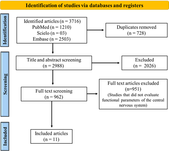
Figure
1 – PRISMA-ScR flow
diagram illustrating the study selection process
Table I – Characteristics
of the included studies
Five studies assessed irisin in healthy
subjects [9,10,11,12,13], reporting conflicting results on its
modulation by exercise. While some studies found increased irisin
levels following aerobic exercise training, others reported no significant
changes, suggesting that exercise intensity and
duration may play a role in its regulation. Reycraft et al. [9] found no significant changes
in irisin concentration after four weeks of aerobic exercise (one session per
week) in older adults. In contrast, Gmiat et al. [10] observed increased irisin
levels following 12 weeks of Nordic walking (three sessions per week) in women.
Rodziewicz-Flis et al. [11] reported
no significant changes in irisin levels after 12 weeks of Nordic walking (three
sessions per week) in healthy older adults but noted significant improvements
in cognitive functions and physical performance. Nicolini et al. [12] found no changes in serum irisin levels in sedentary
but healthy men following an exercise protocol. Meanwhile, Tsai et al. [13] observed increased irisin
levels in healthy older adults only after high-intensity interval training
(HIIT).
Similarly, BDNF was examined in eight studies,
with mixed findings regarding its exercise-induced secretion. Some studies
demonstrated elevated BDNF concentrations post-exercise,
correlating with cognitive improvements, while others found no significant
changes, despite observed enhancements in
cognitive function and neuroprotection. De Poli et al. [14], in a study with physically
active women, found an increase in BDNF levels after a 49-day acute
high-intensity intermittent aerobic exercise protocol (four total sessions). The
study also suggested that peripheral BDNF may reduce tension, depression, and
anger, regardless of menstrual phase. Reycraft et al. [9] reported an intensity-dependent increase in BDNF levels,
associating it with neuroprotection and synaptic plasticity in healthy men. Tsai
et al. [13] reported similar BDNF responses
after high- and moderate-intensity training, linking exercise to improved
neurocognitive performance, though not directly to changes in
myokine concentrations. Gmiat et al. [10] observed increased BDNF levels following exercise in
healthy women, associating irisin and BDNF increases with cognitive
improvement. Nicolini et al. [12] reported
increased serum BDNF levels after HIIT training in healthy participants. Kettinen et al. [15]
found no changes in plasma BDNF levels after two weeks of aerobic exercise
(five sessions per week) in healthy individuals, but reported improvements in
cognitive and executive functions, regardless of changes in myokine levels. Rodziewicz-Flis
et al. [11] also found no changes in
BDNF levels following aerobic exercise. Gaitán et al. [16] observed a decrease in plasma BDNF levels after 26
weeks of aerobic exercise (three sessions per week) in individuals with a
family history of Alzheimer’s disease. However, the same study reported
improvements in cognitive performance.
Of the 11 included studies,
only three
specifically investigated individuals with CNS disorders. Zhang
et al. [17] reported increased irisin
levels following a 12-week multimodal physical training program (52 minutes,
twice per week, comprised muscular strength, balance and aerobic training on
the cycle ergometer) in patients with Parkinson's disease, associating this
increase with improved balance function. Prader-Willi Syndrome patients showed
no significant changes in irisin and BDNF levels after 1 session of resistance
training [18]. Tsai et al. [19]
applied acute aerobic and resistance exercises to men and women with amnesic
mild cognitive impairment, detecting a significant increase in serum BDNF
levels. Improvements in cognitive-behavioral performance were also observed,
assessed via the Flanker test.
Regarding other myokines, one study
reported that aerobic training increased plasma CTSB levels,
associating this with cognitive improvements in healthy subjects with family
history of Alzheimer disease [16]. No significant alterations were found in
IL-6, osteonectin, myostatin, or FGF-21
following exercise interventions in healthy and CNS disorders patients [18].
Tsai et al. [19] reported no
alterations in serum FGF levels but found a significant increase in serum IGF-1
levels following one session of exercise in.
Differences in study populations, sample sizes, and exercise
protocols may account for these inconsistencies reported,
emphasizing the complexity of exercise-myokine-CNS interactions. Table
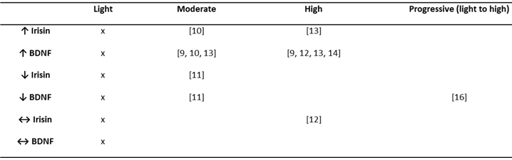
II provides a detailed description of exercise protocols and studies outcomes. Table III summarizes changes in the most studied
myokines (irisin and BDNF) in healthy subjects according to the exercise
protocol.
Table II - Exercise protocol description and myokine results
Table III - Changes in irisin and BDNF induced by aerobic
exercise training in healthy subjects, according to exercise intensity
↑
= increase; ↓ = decrease; ↔ = no change. BDNF: Brain-derived neurotrophic fator. x: no included study applied this exercise intensity
Discussion
The findings of this review
underscore the influence of physical exercise on myokine secretion and its
potential role in CNS function. The most extensively
studied myokines, irisin and BDNF, have been linked to cognitive
improvements, neuroplasticity, and neuroprotection, yet their
secretion patterns appear highly dependent on the specific exercise protocol
intensity. In healthy subjects, aerobic exercise of
moderate or high intensity, regardless of protocol duration, induced increased
secretion of irisin and BDNF. Due to the limited number of studies conducted in
individuals with CNS dysfunctions - and the heterogeneity among them in terms of
diagnoses (Parkinson’s disease, Prader-Willi syndrome, and cognitive decline),
exercise protocols, and outcome measures - it is not possible to draw definitive
conclusions regarding this population.
Studies identified cognitive or mood benefits
of exercise in healthy subjects, despite inconsistent changes
in myokine concentrations. For instance, while six studies [10,11,13,14,15,16]
reported improvements in executive function, memory, mood, and overall
cognitive performance, the association between these effects and irisin and
BDNF levels remained inconclusive. Cognitive and mood improvements associated
with increased myokine levels were observed in all studies that applied
high-intensity aerobic exercise [13,14,16]. It is worth noting that Gaitán et al. [16] conducted a progressive
intensity protocol and did not find an increase in BDNF levels; however, CTSB
levels were associated with cognitive improvements. On the other hand, some
studies reported exercise-induced cognitive improvements despite reductions in
myokine levels [11] or no significant changes [15]. This discrepancy suggests that
exercise intensity may influence these effects, and that exercise can promote
neuroplasticity through mechanisms beyond direct myokine modulation - potentially
involving other neurotrophic factors, inflammatory pathways, or metabolic
adaptations.
Improved cognitive performance
associated with increased myokine levels (BDNF, IGF-1) was reported in older
adults with amnestic mild cognitive impairment following a single session of
aerobic exercise [19]. Beyond cognitive function, other exercise-induced CNS
effects were reported in only one study conducted in patients with Parkinson’s
disease [17]. Irisin levels increased following a multimodal
physical training protocol, and this increase was associated with improved
balance function. Recent evidence suggests that postural control, a motor
symptom, is linked to cognitive function - a non-motor symptom - in Parkinson’s
disease and other neurodegenerative conditions [20,21].
Despite the growing interest in
exercise-induced myokines, several knowledge gaps remain. A key observation
highlighted by our review is that exercise type and intensity may influence
myokine responses. High-intensity training appeared to elicit greater increases
in irisin and BDNF, whereas moderate-intensity aerobic exercise yielded more
variable results. Resistance training, although less frequently studied, also
demonstrated beneficial effects on CNS function - supporting the notion that both
aerobic and resistance exercise contribute to brain health. Consistent with our
findings, recent reviews suggest a dose–response relationship between exercise
intensity and myokine release [22,23]. However, variations in exercise
frequency, intensity, duration, and sample characteristics across studies limit
the ability to draw definitive conclusions. In spite of the relatively small
number of studies, the heterogeneity of findings could be better understood
through systematic reviews and meta-analyses focusing on healthy individuals,
particularly to quantify the effects of exercise intensity on irisin and BDNF
secretion.
Another limitation is that most
studies focused on healthy individuals, restricting our understanding of how
neurological disorders may modulate myokine secretion and function. Only one
study directly compared myokine responses between healthy individuals and those
with CNS dysfunctions, reporting no significant differences [18]. This finding
underscores the need for comparative research to clarify whether CNS
pathologies alter exercise-induced myokine secretion patterns.
Furthermore, most studies assessed
only peripheral myokine levels, which may not accurately reflect their activity
within the CNS. Future investigations should incorporate neuroimaging
techniques, cerebrospinal fluid analysis, and animal models to better elucidate
the mechanisms by which exercise-induced myokines affect brain function.
Conclusion
The findings of this scoping review
highlight the potential of physical exercise, particularly aerobic training at
moderate to high intensities, to modulate myokine secretion in healthy
individuals. Although evidence supporting similar effects in clinical
populations remains scarce and inconclusive, these initial observations support
future investigations aimed at elucidating the role of exercise-induced
myokines as mediators of CNS-related adaptations in both health and disease.
Conflict
of interest
The
authors have no conflicts of interest to declare.
Funding
Statement
This
research received no specific grant from any funding agency in the public,
commercial, or not-for-profit sectors.
Authors'
Contributions
Conceptualization;
Data curation; Formal analysis; Writing – original draft: Neves PHS; Conceptualization; Supervision; Writing – review &
editing: Araújo P
References
- Alves B, Oliveira M. OMS divulga informe
mundial de saúde mental: transformar a saúde mental para todos [Internet].
Biblioteca Virtual em Saúde MS. [cited 2025 Aug 7]. Available
from:
https://bvsms.saude.gov.br/oms-divulga-informe-mundial-de-saude-mental-transformar-a-saude-mental-para-todos/
- Empresa Brasileira de Serviços
Hospitalares. Ações realizadas pela Rede Ebserh/MEC
buscam conscientizar sobre a importância da saúde mental [Internet]. [cited 2025 Aug 7]. Available from: https://www.gov.br/ebserh/pt-br/comunicacao/noticias/acoes-realizadas-pela-rede-ebserh-mec-buscam-conscientizar-sobre-a-importancia-da-saude-mental
- Pedersen
BK. Physical activity and muscle-brain crosstalk. Nat Rev Endocrinol.
2019;15(7):383–92. doi: 10.1038/s41574-019-0174-x [Crossref]
- Febbraio MA, Pedersen BK. Who would have thought – myokines two decades on. Nat Rev Endocrinol. 2020;16(11):619–20. doi: 10.1038/s41574-020-00408-7 [Crossref]
- Bay ML, Pedersen BK. Muscle-organ crosstalk: focus on immunometabolism. Front Physiol. 2020;11:567881. doi: 10.3389/fphys.2020.567881 [Crossref]
- Chow LS, Gerszten RE, Taylor JM, Pedersen BK, van Praag H, Trappe S, et al. Exerkines in health, resilience and disease. Nat Rev Endocrinol. 2022;18(5):273–89. doi: 10.1038/s41574-022-00641-2 [Crossref]
- Young MF, Valaris S, Wrann CD. A role for FNDC5/irisin in the beneficial effects of exercise on the brain and in neurodegenerative diseases. Prog Cardiovasc Dis. 2019;62(2):172–8. doi: 10.1016/j.pcad.2019.02.007 [Crossref]
- Tricco AC, Lillie E, Zarin W, O'Brien KK, Colquhoun H, Levac D, et al. PRISMA extension for scoping reviews (PRISMA-ScR): checklist and explanation. Ann Intern Med. 2018;169(7):467–73. doi: 10.7326/M18-0850 [Crossref]
- Reycraft JT, Islam H, Townsend LK, Hayward GC, Hazell TJ, Macpherson REK. Exercise intensity and recovery on circulating brain-derived neurotrophic factor. Med Sci Sports Exerc. 2020;52(5):1210–7. doi: 10.1249/MSS.0000000000002242 [Crossref]
- Gmiat A, Jaworska J, Micielska K, Kortas J, Prusik K, et al. Improvement of cognitive functions in response to a regular Nordic walking training in elderly women – a change dependent on the training experience. Exp Gerontol. 2018;104:105–12. doi: 10.1016/j.exger.2018.02.006 [Crossref]
- Rodziewicz-Flis E, Juhas U, Kortas JA, Jaworska J, Bidzan-Bluma I, Babińska A, et al. Nordic walking training in BungyPump form improves cognitive functions and induces changes in amino acids and kynurenine profiles in older adults. Front Endocrinol (Lausanne). 2023;14:123456. doi: 10.3389/fendo.2023.1151184 [Crossref]
- Nicolini C, Michalski B, Toepp SL, Turco CV, D'Hoine T, Harasym D, et al. A single bout of high-intensity interval exercise increases corticospinal excitability and BDNF in sedentary healthy males. Brain Res. 2020;437:242–55. doi: 10.1016/j.neuroscience.2020.03.042 [Crossref]
- Tsai CL, Pan CY, Tseng YT, Chen FC, Chang YC, Wang TC. Acute effects of high-intensity interval training and moderate-intensity continuous exercise on BDNF and irisin levels and neurocognitive performance in late middle-aged and older adults. Behav Brain Res. 2021;413:113472. doi: 10.1016/j.bbr.2021.113472 [Crossref]
- De Poli RAB, Lopes VHF, Lira FS, Zagatto AM, Jimenez-Maldonato A, Antunes BM. Peripheral BDNF and psycho-behavioral aspects are positively modulated by high-intensity intermittent exercise and fitness in healthy women. Sci Rep. 2021;11(1):3810. doi: 10.1038/s41598-021-83072-9 [Crossref]
- and biomarker responses in healthy older adults to an 18-hole golf round and different walking types: a randomised cross-over study. BMJ Open Sport Exerc Med. 2023;9(4):e001629. doi: 10.1136/bmjsem-2023-001629 [Crossref]
- Gaitán JM, Moon HY, Stremlau M, Dubal DB, Cook DB, Okonkwo OC, et al. Effects of aerobic exercise training on systemic biomarkers and cognition in late middle-aged adults at risk for Alzheimer’s disease. Front Endocrinol (Lausanne). 2021;12:660546. doi: 10.3389/fendo.2021.660181 [Crossref]
- Zhang X, Xu S, Hu Y, Liu Q, Liu C, Chai H, et al. Irisin exhibits neuroprotection by preventing mitochondrial damage in Parkinson’s disease. NPJ Parkinsons Dis. 2023;9(1):7. doi: 10.1038/s41531-023-00453-9 [Crossref]
- Hirsch HJ, Gross-Tsur V, Sabag Y, Nice S, Genstil L, Benarroch F, et al. Myokine levels after resistance exercise in young adults with Prader–Willi syndrome (PWS). Am J Med Genet A. 2020;182(1):115–21. doi: 10.1002/ajmg.a.61391 [Crossref]
- Tsai CL, Ukropec J, Ukropcová B, Pai MC. An acute bout of aerobic or strength exercise specifically modifies circulating exerkine levels and neurocognitive functions in elderly individuals with mild cognitive impairment. Neuroimage Clin. 2017;17:272–84. doi: 10.1016/j.nicl.2017.10.028 [Crossref]
- Tait P, Graham L, Vitorio R, Watermeyer T, Timm EC, O'Keefe J, et al. Neuroimaging and cognitive correlates of postural control in Parkinson’s disease: a systematic review. J Neuroeng Rehabil. 2025;22:24. doi: 10.1186/s12984-024-01539-y [Crossref]
- Bae M, Zheng P, VanNostrand M. The effects of balance training on cognitive function in persons with multiple sclerosis: a systematic review and meta-analysis. Mult Scler Relat Disord. 2025;94:106274. doi: 10.1016/j.msard.2025.106274 [Crossref]
- Zare N, Bishop DJ, Levinger I, Febbraio MA, Broatch JR. Exercise intensity matters: a review on evaluating the effects of aerobic exercise intensity on muscle-derived neuroprotective myokines. Alzheimers Dement (N Y). 2025;11(1):e70056. doi: 10.1002/trc2.70056 [Crossref]
- Gao X, Chen Y, Cheng P. Unlocking the potential of exercise: harnessing myokines to delay musculoskeletal aging and improve cognitive health. Front Physiol. 2024;15:1338875. doi: 10.3389/fphys.2024.1338875 [Crossref]
