Rev Bras Fisiol Exerc. 2026;25(2):e255667
doi: 10.33233/rbfex.v25i2.5667ORIGINAL ARTICLE
Carotid morphofunctional
responses to maximal aerobic
exercise in patients with coronary artery
disease
Respostas
morfofuncionais da carótida ao exercício aeróbio máximo em pacientes com doença
arterial coronariana
Gabriela Andrade Paz1,
André Luiz Conveniente Soares1, Tatiana Rodrigues dos Santos1,
Caio Luan Farias Costa1, Cláudia de Mello Meirelles1,2,
Paulo Sergio Chagas Gomes1, Maria das Graças Coelho de Souza1,
Juliana Pereira Borges1
1Universidade do Estado do Rio de
Janeiro, Rio de Janeiro, RJ, Brasil
2Escola de Educação Física do Exército (EsEFEx), Rio de Janeiro, RJ, Brasil
Received: March 13,
2026; Accepted: April 17, 2026.
Correspondence: Gabriela Andrade Paz, gabrielapaz.nutri@gmail.com
How to cite
Paz GA, Soares ALC, Santos TR,
Costa CLF, Meirelles CM, Gomes PSC, Souza MGC, Borges JP. Carotid
morphofunctional responses to
maximal aerobic exercise in
patients with coronary artery disease. Rev Bras
Fisiol Exerc
2026;25(2):e255667. doi: 10.33233/rbfex.v25i2.5667
Abstract
Objective: To evaluate the acute effects of maximal aerobic
exercise on carotid morphofunctional markers in patients with coronary artery
disease (CAD) and healthy individuals. Methods: Patients with CAD (n =
8; 62.3 ± 4.3 years; 27.1 ± 3.1 kg/m²) who had completed phase II of a cardiac
rehabilitation program (CRP) and healthy individuals (CONT; n = 5; 61.6 ± 3.5
years; 27.3 ± 3.2 kg/m²) were evaluated for carotid morphofunctional markers by
ultrasonography before and after a cardiopulmonary exercise test (CPET). Results:
Maximal oxygen uptake was lower in the CAD group compared with CONT (16.7 ± 3.5
vs. 24.0 ± 5.2 mL•kg-¹•min-¹; P = 0.01). Before CPET, no
differences were observed between groups for systolic diameter (CAD: 6.05±0.65
vs. CONT: 6.23 ± 0.20 mm; P = 0.6), diastolic diameter (CAD: 5.41 ± 0.73 vs.
CONT: 5.73 ± 0.15 mm; P = 0.4), peak systolic velocity (CAD: 66.27 ± 12.24 vs.
CONT: 61.33± 15.67 cm•s-¹; P = 0.4), or end-diastolic velocity (CAD:
20.63 ± 4.15 vs. CONT: 21.07 ± 3.97 cm•s-¹; P = 0.4). Carotid
intima–media thickness (cIMT) was borderline higher in the CAD group (0.93 ±
0.23 vs. 0.71 ± 0.10 mm; P = 0.07). After CPET, both groups showed increases in
systolic diameter (CAD: +0.26; CONT: +0.16 mm; P < 0.001), diastolic diameter
(CAD: +0.31; CONT: +0.17 mm; P = 0.03), and peak systolic velocity (CAD: +
9.80; CONT: + 8.66 cm·s-¹; P = 0.04). End-diastolic velocity (CAD: +
0.60; CONT: -3.75 cm·s-¹; P = 0.4) and cIMT (CAD: - 0.02; CONT: -0.01
mm; P = 0.1) did not change significantly in either group (P > 0.05). No
between-group differences were observed in CPET-induced changes. Conclusion:
Patients with CAD who had completed phase II of a cardiac rehabilitation
program exhibited preserved vascular responses to maximal exercise.
Keywords: physical exercise; coronary artery disease;
atherosclerosis; endothelial dysfunction
Resumo
Objetivo: Avaliar os efeitos agudos do exercício
aeróbio máximo sobre marcadores morfofuncionais da carótida em pacientes com
doença arterial coronariana (DAC) e indivíduos saudáveis. Métodos:
Pacientes com DAC (n = 8; 62,3 ± 4,3 anos; 27,1 ± 3,1 kg/m²), que completaram a
fase II de um programa de reabilitação cardíaca (PRC), e indivíduos saudáveis
(CONT; n = 5; 61,6 ± 3,5 anos; 27,3 ± 3,2 kg/m²) foram avaliados quanto aos
marcadores morfofuncionais da carótida por ultrassonografia antes e após um
teste cardiopulmonar de exercício (TCPE). Resultados: O consumo máximo
de oxigênio foi menor em DAC em comparação com CONT (16,7 ± 3,5 vs. 24,0 ± 5,2
ml/kg/min; P = 0,01). Antes do TCPE, não foram observadas diferenças entre os
grupos para diâmetro sistólico (DAC: 6,05 ± 0,65 vs. CONT: 6,23 ± 0,20 mm; P =
0,6), diâmetro diastólico (DAC: 5,41 ± 0,73 vs. CONT: 5,73 ± 0,15 mm; P = 0,4),
velocidade de pico sistólico (DAC: 66,27 ± 12,24 vs. CONT: 61,33 ± 15,67
cm.s-1; P = 0,4) e velocidade diastólica final (DAC: 20,63 ± 4,15 vs. CONT:
21,07 ± 3,97 cm.s-1; P=0,4). A espessura íntima-média carotídea (cIMT) apresentou valor limítrofe mais elevado em DAC (0,93
± 0,23 vs. 0,71 ± 0,10 mm; P = 0,07). Após o TCPE, ambos os grupos aumentaram o
diâmetro sistólico (DAC: +0,26; CONT: + 0,16 mm; P < 0,001), diâmetro
diastólico (DAC: + 0,31; CONT: + 0,17 mm; P = 0,03) e velocidade de pico
sistólico (DAC: + 4,18; CONT: + 8,66 cm.s-1; P = 0,01). A velocidade diastólica
final (DAC: -2,05; CONT: -3,75 cm.s-1; P = 0,5) e a cIMT
(DAC: -0,02; CONT: -0,01 mm; P = 0,1) não se alteraram significativamente em
nenhum grupo (P > 0,05). Não foram observadas diferenças entre os grupos nas
alterações induzidas pelo TCPE. Conclusão: Pacientes com DAC que
concluíram a fase II do PRC apresentaram respostas vasculares preservadas ao
exercício máximo.
Palavras-chave: exercício físico; doença arterial
coronariana; aterosclerose; disfunção endotelial
Introduction
Coronary artery disease (CAD) is one of the leading
causes of morbidity and mortality worldwide among cardiovascular diseases [1].
Traditional risk factors, such as diabetes, hypertension, dyslipidemia, and
physical inactivity, significantly contribute to its development [2]. The CAD
is the main clinical manifestation of atherosclerosis, a condition
characterized by the progressive accumulation of lipid plaques in the walls of
medium and large-sized arteries, impairing blood flow and potentially leading
to severe cardiovascular events [3,4].
In addition to arterial obstruction, the chronic
inflammatory process inherent to atherosclerosis and the resulting impairment
of endothelial function are associated with systemic vascular dysfunction,
including morphofunctional alterations of the carotid artery. Among these
alterations, increases in carotid intima-media thickness (cIMT) stand out,
along with changes in systolic and diastolic diameters, peak systolic velocity,
and end-diastolic velocity [4,5,6].
These markers are widely used to assess arterial
structure, blood flow, and hemodynamic changes, as well as to identify
atherosclerotic plaques and significant stenosis. The cIMT is considered a
subclinical biomarker of atherosclerosis, whereas peak systolic velocity is
frequently used to detect hemodynamically relevant stenosis. These hemodynamic
characteristics can be assessed using B-mode ultrasound with Doppler, and
alterations in these markers are recognized as independent predictors of
cardiovascular risk [7].
Evidence indicates that regular practice of multimodal
physical exercise, including aerobic, resistance, and flexibility exercises,
reduces the risk of cardiovascular complications [8]. In particular, aerobic
training may induce vascular adaptations, with studies demonstrating benefits
on cIMT in different populations, including healthy individuals [9,10].
Maximal aerobic exercise can be assessed during
cardiopulmonary exercise testing (CPET), which allows the determination of
maximal oxygen uptake (VO2max), an important indicator of
cardiorespiratory fitness. The CPET consists of a progressive, individualized
protocol that gradually increases workload until maximal effort is reached,
enabling the integrated analysis of cardiovascular, respiratory, and metabolic
responses to exercise. Changes in vascular function, such as those observed in
atherosclerosis, may be associated with reduced CPET performance and lower VO2max
[11,12]. Despite the recognized benefits of physical training, studies
investigating the acute responses of carotid morphofunctional markers to
maximal aerobic exercise in patients with CAD are still scarce, especially
following structured cardiac rehabilitation programs. Therefore, the aim of the
present study was to evaluate the impact of maximal aerobic exercise on carotid
morphofunctional markers in patients with CAD participating in a cardiac
rehabilitation program and in healthy individuals, considering that maximal
exercise induced by CPET may reveal relevant vascular responses in this
population.
Methods
Participants
Eight patients with CAD were recruited from the Hospital
Universitário Pedro Ernesto (HUPE), and five healthy volunteers without
evidence of CAD (CONT) were randomly recruited. Both groups included
individuals of both sexes aged 50-70 years with a body mass index (BMI) < 30
kg/m². Due to the exploratory nature of the study and the availability of
eligible patients during the data collection period, no formal sample size
calculation was performed.
Patients with CAD were considered eligible if they had
completed phase II of the HUPE cardiac rehabilitation program, which included
individualized physical exercise, including both aerobic and resistance
training, varying according to the patient’s capacity, generally between 50%
and 60% of maximum heart rate. CAD was defined as the presence of acute
coronary syndrome (myocardial infarction with or without ST-segment elevation
or unstable angina) or obstructive CAD, diagnosed by coronary angiography, in
patients with stable angina.
Exclusion criteria for the CAD group included: a)
diagnosis of heart failure, pulmonary disease, Chagas disease, or tuberculosis;
b) regular physical exercise practice (≥ 3 days/week for 30 min) in the last 6 months; c)
malnutrition (BMI < 18.5 kg/m²); d) smoking; and e) obesity (BMI < 30 kg/m²). Individuals in the CONT group were also
excluded if they had a previous or current diagnosis of hypertension, ischemic
disease, and/or diabetes mellitus, in addition to having a pacemaker or using
antidepressant medication.
All participants received oral and written instructions
regarding the risks and benefits of the study and signed an informed consent
form. The study was registered at ClinicalTrials.gov (NCT06579807), approved by
the HUPE Research Ethics Committee (CAAE 81718324.3.0000.5259), and conducted
in accordance with the SPIRIT 2013 recommendations (Standard Protocol Items:
Recommendations for Interventional Trials) [13].
Study design
This is an observational study with repeated measures,
with assessments performed before and immediately after maximal exercise. The
evaluations were conducted during a single visit to the Laboratory of Physical
Activity and Health Promotion of the State University of Rio de Janeiro, always
in the morning (8:00–11:00 a.m.), in a room with controlled temperature between
22–24°C and relative humidity between 60–70%. Participants were instructed to
avoid physical activity, alcohol, and stimulants for 48 hours prior to the
assessments.
Initially, medical history
was assessed. After 20
minutes of rest in the supine position, a carotid ultrasound evaluation was
performed. Subsequently, blood pressure (BP) was measured using an automatic
digital device (Omron™, Matsusaka, Japan), along with resting heart rate (HR),
prior to CPET. Thereafter, CPET was performed, and within 5 minutes after its
completion, carotid ultrasound evaluation was repeated. Figure 1 illustrates
the study's experimental design.
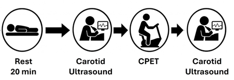
Figure 1 - Experimental design. CPET = cardiopulmonary
exercise testing
Carotid artery morphofunctional assessment
Carotid intima-media thickness (cIMT) of the right common
carotid artery was assessed with the participant in the supine position, with
the head turned to the side (45°), using a portable ultrasound device (GE
LOGIQ, General Electric Healthcare, Chicago, IL, USA) equipped with a 12L-RS
linear probe (38 mm) operating at a frequency of 10 MHz.
Real-time B-mode ultrasound enabled longitudinal carotid
artery imaging to confirm cIMT measurements. All measurements were performed by
a trained technician and were taken 10 to 20 mm proximal to the carotid
bifurcation (carotid bulb), focusing on the far wall, which provides better
definition [7]. The cIMT values were manually measured as the mean of three
measurements using ImageJ (NIH, USA).
B-mode Doppler ultrasound also allowed assessment of the
systolic (SD) and diastolic (DD) diameters of the carotid artery. From Doppler
analysis, peak systolic velocity (PSV) and end-diastolic velocity (EDV) were
obtained using the Triplex ultrasound technique, which combines B-mode imaging
with color Doppler and pulsed-wave Doppler spectral analysis. To improve
velocity measurement accuracy, the insonation angle was set between 30° and 60°
[7].
Cardiopulmonary exercise testing
The test was performed on an electromagnetically braked
cycle ergometer (Inbramed®, Porto Alegre, Brazil) connected to a computer,
using an individualized ramp protocol. The protocol was developed based on
estimated VO2max values using a non-exercise model [14]. The
cardiorespiratory fitness model was designed for individuals aged 18 to 80
years and can be applied to both men and women. Based on these values, the
initial and final workload rates and the workload increment rates were
calculated [15] using the metabolic equations proposed by the American College
of Sports Medicine [16] to ensure the test duration ranged from 8 to 12
minutes.
During the CPET, gas exchange and ventilatory variables
(VO2, VCO2, and VE) were collected breath-by-breath using
a silicone mask (Hans Rudolph®, Shawnee, USA) connected to an Ultima CardiO2
metabolic cart (Medical Graphics®, Saint Louis, USA) [17]. Gas analyzers were
calibrated before each test according to the manufacturer’s instructions, using
a certified standard gas mixture containing oxygen (17.01%) and carbon dioxide
(5.00%), balanced with nitrogen (AGAR, Rio de Janeiro, RJ, Brazil). The
accuracy of flow and volume measurements obtained by the pneumotachograph was verified
using a 3-L calibration syringe (Hans Rudolph®, Kansas, USA).
Data were averaged over 30-s intervals, and VO2max
was defined as the highest 30-s average obtained during the test. For the test
to be considered maximal, participants were required to meet at least three of
the following criteria [18]: a) volitional exhaustion; b) achievement of 90% of
age-predicted maximal heart rate (HR) estimated by the equation 220 - age or
absence of HR increase with increasing workload; c) presence of a VO2
plateau (variation < 2.1 ml·kg-¹·min-¹ between two
consecutive workloads); d) respiratory exchange ratio (R) > 1.1; and e) Borg
scale rating ≥ 17.
Statistical analysis
Data were averaged over 30-s intervals, and VO2max
was defined as the highest 30-s average obtained during the test. For the test
to be considered maximal, participants were required to meet at least three of
the following criteria [18]: a) volitional exhaustion; b) achievement of 90% of
age-predicted maximal heart rate (HR) estimated by the equation 220 - age or
absence of HR increase with increasing workload; c) presence of a VO2
plateau (variation < 2.1 ml·kg-¹·min-¹ between two
consecutive workloads); d) respiratory exchange ratio (R) > 1.1; and e) Borg
scale rating ≥ 17.
Results
Clinical and cardiorespiratory characteristics of the
participants are presented in Table I. No significant differences were observed
between the CAD and CONT groups regarding sex distribution, age, body mass
index, resting systolic and diastolic blood pressure, or resting heart rate. In
contrast, VO2max was significantly lower in the CAD group compared
to the CONT group (P = 0.01). Patients in the CAD group were using statins
(87.5%), antiplatelet agents (75%), beta-blockers (62.5%), and
renin–angiotensin–aldosterone system inhibitors (75%). In the CONT group, only
20% of the volunteers were using statins, with no use of medications from other
classes.
Table I - Clinical characteristics of the CAD and CONT groups
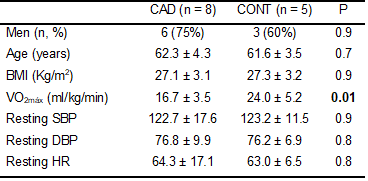
CAD = coronary artery disease patients; CONT = healthy
individuals; VO2max = maximal oxygen uptake; SBP = systolic blood
pressure; DBP = diastolic blood pressure; HR = heart rate
The carotid artery ultrasonographic variables assessed
before and immediately after CPET are described in Table II. At pre-CPET, no
differences were observed between groups in systolic and diastolic diameters,
peak systolic velocity, or end-diastolic velocity. The cIMT was borderline
higher in the CAD group than in the CONT group (P = 0.07).
After CPET, a similar increase was observed in both
groups for carotid systolic and diastolic diameters, peak systolic velocity,
and heart rate, with no differences between groups. End-diastolic velocity and
cIMT did not show significant changes after CPET in either group.
Table II - Mean (standard deviation) of carotid
ultrasonographic variables pre- and post-CPET in patients with coronary artery
disease (CAD) and healthy individuals (CONT)
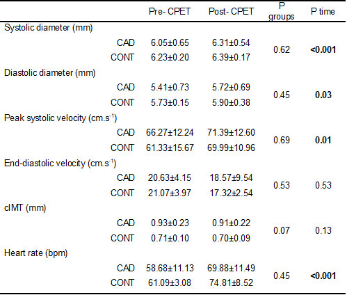
CPET
= cardiopulmonary exercise testing; cIMT = carotid intima-media thickness
.
Absolute changes (post–pre-CPET) in carotid
ultrasonographic variables are presented in Figure 2. No significant
differences were observed between the CAD and CONT groups in maximal
exercise-induced responses for any of the evaluated variables.
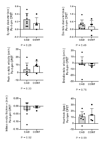
Figure 2 - Changes in carotid ultrasonographic variables with
maximal aerobic exercise
Discussion
The main finding of the present study was that patients
with CAD, after phase II of a cardiac rehabilitation program, exhibited carotid
artery morphofunctional responses similar to those observed in healthy
individuals during maximal aerobic exercise. Despite the lower VO2max
observed in the CAD group, the acute changes induced by CPET, characterized by
increases in systolic and diastolic diameters and peak systolic velocity,
occurred similarly between groups, suggesting preserved vascular responsiveness
in this population.
The lower cardiorespiratory fitness observed in patients
with CAD is consistent with previous evidence associating cardiovascular
impairment and atherosclerosis with reduced VO2max [12]. Studies
have shown that structural and functional alterations of the cardiovascular
system, including endothelial dysfunction and increased arterial stiffness, are
associated with poorer cardiorespiratory performance [19,20]. However, our
findings indicate that, despite overall functional impairment, the carotid
artery's acute response to maximal exercise may be preserved in clinically
stable patients participating in cardiac rehabilitation programs.
The cIMT showed borderline higher values in the CAD group
than in the CONT group, although this difference was not statistically
significant. The absence of changes in cIMT after CPET was expected, as this
marker reflects chronic structural adaptations of the arterial wall and is not
readily influenced by acute changes induced by a single exercise session.
Consistent evidence indicates that significant reductions in cIMT occur
predominantly after long-term exercise interventions [9], generally exceeding
six months, especially in individuals with elevated baseline values or
atherosclerotic plaques [10]. Thus, the absence of cIMT changes in the present
study does not negate the beneficial effects of exercise but reinforces the
distinction between acute vascular adaptations and long-term structural
remodeling.
On the other hand, the increases observed in carotid
systolic and diastolic diameters, as well as in peak systolic velocity after
the CPET, reflect acute hemodynamic adaptations to maximal exercise. These
responses are associated with increases in cardiac output, pulse pressure, and
arterial wall shear stress, which stimulate endothelium-dependent vasodilation
mechanisms, as described in the literature [21,22]. The preservation of these
responses in the CAD group suggests maintenance of vascular accommodation
capacity during transient increases in hemodynamic demand, possibly favored by
prior participation in a structured cardiac rehabilitation program.
Although parameters such as flow velocity and arterial
diameter are widely used to assess vascular function, there are few studies
specifically investigating their acute responses to maximal aerobic exercise in
patients with CAD.
Previous studies in healthy individuals have shown that
acute exercise may temporarily increase arterial stiffness and modify carotid
hemodynamic parameters. Liu et al. [23] observed acute increases in elastic
modulus and arterial diameter immediately after cycling exercise, with
progressive attenuation of these responses over weeks of training. Similarly,
Yuan et al. [24] reported a transient increase in carotid stiffness and
elevation of the oscillatory shear index after high-intensity interval exercise.
These changes are mainly attributed to abrupt increases in systolic blood
pressure, heart rate, and pulsatile flow, which elevate shear stress and
circumferential wall tension. In the present study, although arterial stiffness
indices were not directly assessed, the increases observed in systolic and
diastolic diameters and peak systolic velocity are consistent with an acute
hemodynamic response to increased cardiac output and pulse pressure, as
described in healthy individuals. Thus, the findings of the present study help
fill this gap, indicating that maximal exercise is well tolerated by clinically
stable patients after phase II of cardiac rehabilitation and can elicit
vascular responses comparable to those observed in individuals without
cardiovascular disease.
Some limitations should be considered. The main
limitation is the small sample size, which limits the generalizability of the
findings and prevents stratified analyses, such as comparisons between patients
with and without atherosclerotic plaques. The absence of significant
differences between groups should be interpreted with caution, considering the
small sample size and the consequent low statistical power. In addition,
ultrasonographic assessment was restricted to the right common carotid artery,
without bilateral analysis or evaluation of other vascular territories.
Although the common carotid artery is widely used as a representative site for
central vascular alterations, this approach limits broader inferences about
systemic circulation and may mask heterogeneous responses across vascular beds.
Future studies, including bilateral carotid assessment, larger samples, and
stratification by degree of carotid atherosclerosis, are needed to further
elucidate vascular responses to exercise in this population.
Conclusion
Maximal aerobic exercise increased carotid diameter and
peak systolic velocity similarly in both patients with CAD and healthy
individuals, with no changes in cIMT or end-diastolic velocity after CPET.
Overall, the results suggest that, in patients with stable CAD after phase II
of cardiac rehabilitation, the acute carotid vascular response to maximal
exercise is preserved compared with healthy individuals, despite lower
cardiorespiratory fitness.
Author affiliations
This article represents part of the doctoral thesis of
Gabriela Andrade Paz, supervised by Prof. Dr. Juliana Pereira Borges and Prof.
Dr. Maria das Graças Coelho de Souza at the University of Rio de Janeiro
State.
Conflict of interests
The authors declare no conflict of interest.
Funding
This study was partially funded by grants from the
National Council for Scientific and Technological Development (CNPq) (grant
numbers: 305332/2024-4 and 404204/2023-6, beneficiary JB) and from the Carlos
Chagas Filho Foundation for Research Support of the State of Rio de Janeiro
(FAPERJ) (grant number: E-26/204.202/2025, beneficiary JB).
Author contributions
Concept and study design:
Paz GA, Souza MGC, Borges JP; Data acquisition: Paz GA, Soares ALC,
Santos TR, Costa CLF; Data analysis and interpretation: Paz GA, Soares
ALC, Borges JP; Statistical analysis: Paz GA, Borges JP; Funding acquisition:
Souza MGC, Borges JP; Manuscript writing: Paz GA, Souza MGC, Borges JP; Critical
revision of the manuscript for important intellectual content: Paz GA,
Soares ALC, Santos TR, Costa CLF, Meirelles CM, Gomes PSC, Souza MGC, Borges
JP.
References
- Tan SCW, Zheng BB, Tang ML, Chu H, Zhao YT, Weng C. Global burden of
cardiovascular diseases and its risk factors, 1990-2021: a systematic analysis
for the global burden of disease study 2021. QJM. 2025;118(6):411-22. doi: 10.1093/qjmed/hcaf022 [Crossref]
- Libby P, Buring JE, Badimon L, Hansson GK, Deanfield J, Bittencourt MS, et al. Atherosclerosis. Nat Rev Dis Primers. 2019;5(1):56. doi: 10.1038/s41572-019-0106-z [Crossref]
- Baumgartner R, Forteza MJ, Ketelhuth DFJ. The interplay between cytokines and the Kynurenine pathway in inflammation and atherosclerosis. Cytokine. 2019;122:154148.. doi: 10.1016/j.cyto.2017.09.004 [Crossref]
- Jebari-Benslaiman S, Galicia-García U, Larrea-Sebal A, Olaetxea JR, Alloza I, Vandenbroeck K, et al. Pathophysiology of atherosclerosis. Int J Mol Sci. 2022;23(6):3346. doi: 10.3390/ijms23063346 [Crossref]
- Bentzon JF, Otsuka F, Virmani R, Falk E. Mechanisms of plaque formation and rupture. Circ Res. 2014;114(12):1852-66. doi: 10.1161/CIRCRESAHA.114.302721 [Crossref]
- Libby P. The changing landscape of atherosclerosis. Nature. 2021;592(7855):524-33. doi: 110.1038/s41586-021-03392-8 [Crossref]
- Lee W. General principles of carotid Doppler ultrasonography. Ultrasonography. 2014;33(1):11-7. doi: 10.14366/usg.13018 [Crossref]
- Carvalho T, Milani M, Ferraz AS, Silveira ADD, Herdy AH, Hossri CAC, et al. Brazilian Cardiovascular Rehabilitation Guideline - 2020. Arq Bras Cardiol. 2020;114(5):943-87. doi: 10.36660/abc.20200407 [Crossref]
- Byrkjeland R, Stensaeth KH, Anderssen S, Njerve IU, Arnesen H, Seljeflot I, et al. Effects of exercise training on carotid intima-media thickness in patients with type 2 diabetes and coronary artery disease. Influence of carotid plaques. Cardiovasc Diabetol. 2016;15:13. doi: 10.1186/s12933-016-0336-2 [Crossref]
- Wang Y, Wu H, Sun J, Wei M, Wang J, Li H, et al. Effect of exercise on carotid artery intima–media thickness in adults: A systematic review and meta-analysis. J Phys Act Health. 2022;19(12):855-67. doi: 10.1123/jpah.2022-0372 [Crossref]
- Nesti L, Santoni L, Frascerra S, Chiriacò M, Natali AJIJoSN, Metabolism E.
Comparison of ramp and step graded cardiopulmonary exercise tests for the
estimate of substrate oxidation rates in recreationally active individuals. Int
J Sport Nutr Exerc Metab. 2025 May 30;35(5):394-403. doi: 10.1123/ijsnem.2024-0236 [Crossref]
- Lind L, Michaëlsson KJCP, Imaging F. Detailed investigation of multiple resting cardiovascular parameters in relation to physical fitness. Clin Physiol Funct Imaging. 2023;43(2):120-7. doi: 10.1111/cpf.12800 [Crossref]
- Chan AW, Tetzlaff JM, Gotzsche PC, Altman DG, Mann H, Berlin JA, et al. SPIRIT 2013 explanation and elaboration: guidance for protocols of clinical trials. BMJ. 2013;346:e7586. doi: 10.1136/bmj.e7586 [Crossref]
- Matthews CE, Heil DP, Freedson PS, Pastides H. Classification of
cardiorespiratory fitness without exercise testing. Medicine and science in
sports and exercise. Med Sci Sports Exerc. 1999;31(3):486-93. doi: 10.1097/00005768-199903000-00019 [Crossref]
- Cunha F, Midgley A, Montenegro R, Vasconcellos F, Farinatti PJIjosm. Utility of a non-exercise VO2max prediction model for designing ramp test protocols. Int J Sports Med. 2015;36(10):796-802. doi: 10.1055/s-0034-1395590 [Crossref]
- Pescatello LS. ACSM's guidelines
for exercise testing and prescription. Philadelphia: Lippincott Williams &
Wilkins; 2013
- Michalski AC, Ferreira AS, Midgley AW, Costa VAB, Fonseca GF, da Silva NSL,
et al. Mixed circuit training acutely reduces arterial stiffness in patients
with chronic stroke: a crossover randomized controlled trial. Eur J Appl
Physiol. 2023;123(1):121-34. doi: 10.1007/s00421-022-05061-8 [Crossref]
- Howley ET, Bassett DR, Welch HG. Criteria for maximal oxygen uptake: review
and commentary. Med Sci Sports Exerc. 1995
Sep;27(9):1292-301. PMID: 8531628
- Größer V, Weyh C, Böttrich T, Frech T, Nolte S, Sommer N, et al. Association of cardiorespiratory fitness level with vascular function and subclinical atherosclerosis in the elderly. Eur J Appl Physiol. 2024;124(5):1487-97. doi: 10.1007/s00421-023-05375-1 [Crossref]
- Nayor M, Gajjar P, Miller P, Murthy VL, Shah RV, Houstis NE, et al. Arterial stiffness and cardiorespiratory fitness impairment in the community. Heart Assoc. 2023;12(21):e029619. doi: 10.1161/JAHA.123.029619 [Crossref]
- Green DJ, Hopman MT, Padilla J, Laughlin MH, Thijssen DHJPr. Vascular
adaptation to exercise in humans: role of hemodynamic stimuli. Physiol Rev.
2017;97(2):495-528. doi: 10.1152/physrev.00014.2016 [Crossref]
- Thijssen DH, Bruno RM, van Mil AC, Holder SM, Faita F, Greyling A, et al. Expert consensus and evidence-based recommendations for the assessment of flow-mediated dilation in humans. Eur Heart J. 2019;40(30):2534-47. doi: 10.1093/eurheartj/ehz350 [Crossref]
- Liu HB, Yuan WX, Wang QY, Wang YX, Cao HW, Xu J, et al. Carotid arterial stiffness and hemodynamic responses to acute cycling intervention at different times during 12-week supervised exercise training period. Biomed Res Int. 2018;2018:2907548. doi: 10.1155/2018/2907548 [Crossref]
- Yuan W, Liu H, Luan Z, Zhao Z, Shen B. Acute effect of high-intensity interval cycling on carotid arterial stiffness and hemodynamics. Biomed Res Int. 2019:2019:6260286. doi: 10.1155/2019/6260286 [Crossref]
